Antibiotics: an ongoing war (part 4)
In the laboratory, we can measure the effectiveness of an antibiotic through various tests that let us understand which is more effective against a given pathogen so we can develop the right therapy.
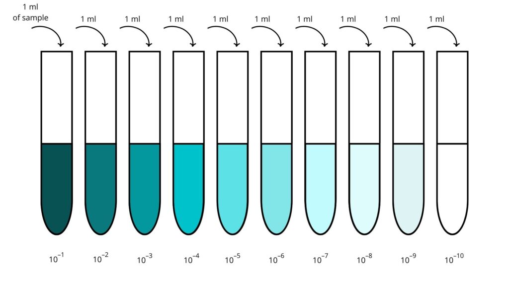
Fundamental to determining the MLC and MIC values is the so-called dilution test. The steps are the following. Firstly, we need to take ten sterile test tubes containing culture medium, a sugar and protein-rich substance bacteria need to grow and replicate. Then we add a quantity of the antibiotic in increasing concentration to nine of these (i.e. an ever-increasing amount of antibiotic). The antibiotic-free vial is needed to verify that bacteria grow correctly without the drug. After that, an equal quantity of bacteria is added in each phial. Finally, they are all placed in a chamber at the temperature of 37°C for about 24 hours (a procedure named incubation). On the following day, we will observe the test tubes. The turbidity indicates the presence of bacteria: it will be maximum in the tube without the antibiotic and will decrease going towards the test tubes with the most antibiotic. The concentration of the first test tube in increasing order in which the medium is transparent indicates the MIC.
On the other hand, determining MLC involves taking the bacteria from the transparent test tubes and placing them inside Petri dishes, which also contain the culture medium (a process called plating). Each capsule will have the same number as the original tube of the contained bacteria: the first capsule free of bacteria in ascending order will indicate the tube whose concentration corresponds to the MLC.
Although these may initially seem complex concepts, let us try to explain them more simply: the greater the concentration of antibiotic used against the bacteria, the higher its toxicity towards them. A low concentration of antibiotics (of the order of µg, i.e. 10^-6 grams) will have no toxic effect on the bacteria. By gradually increasing the concentration of the drug, its action will get more and more effective. Once we have reached a specific concentration, the bacteria will no longer be able to replicate, remaining still alive. By adding more antibiotics, the cells will eventually die. The first of the two values is the MIC, while the second corresponds to the MLC.
One way to understand a bacterium’s susceptibility to various antibiotics is the Kirby-Bauer test. Devised by William Kirby and AW Bauer in the 1960s, it is widely used and is also one of the simplest. The test starts with placing the bacterium of interest in a Petri dish. After that, some tablets containing different antibiotics are placed onto the dish at equal distances from each other. The plate is incubated at 37°C for about 24 hours and then observed.
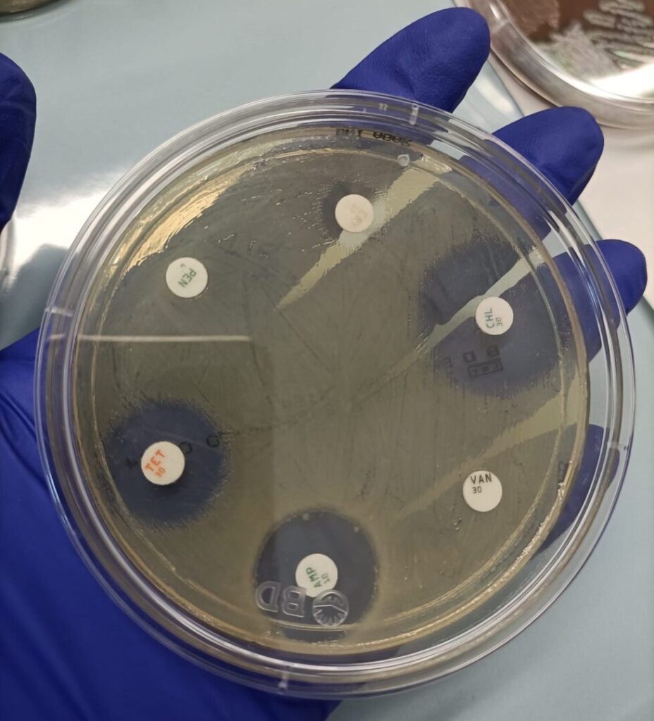
As shown in the image, bacteria have normally grown around some lozenges: in that case, the bacteria are resistant to the antibiotic. At the same time, there are halos around some other tablets: this time bacteria are more or less susceptible to the antibiotic since they have not grown (something like what happened to Fleming with mould). This test can help identify bacterial species, too. Measuring the diameter of the halos, we can indeed calculate the grade of susceptibility of the organism toward each drug, which is generally species-specific.